Article with drawings and photographs from the Illustrated London News showing developments in X-rays used to detect bullets in wounded soldiers, 6 February 1915, (Catalogue ref: ZPER 34/147)
Transcript
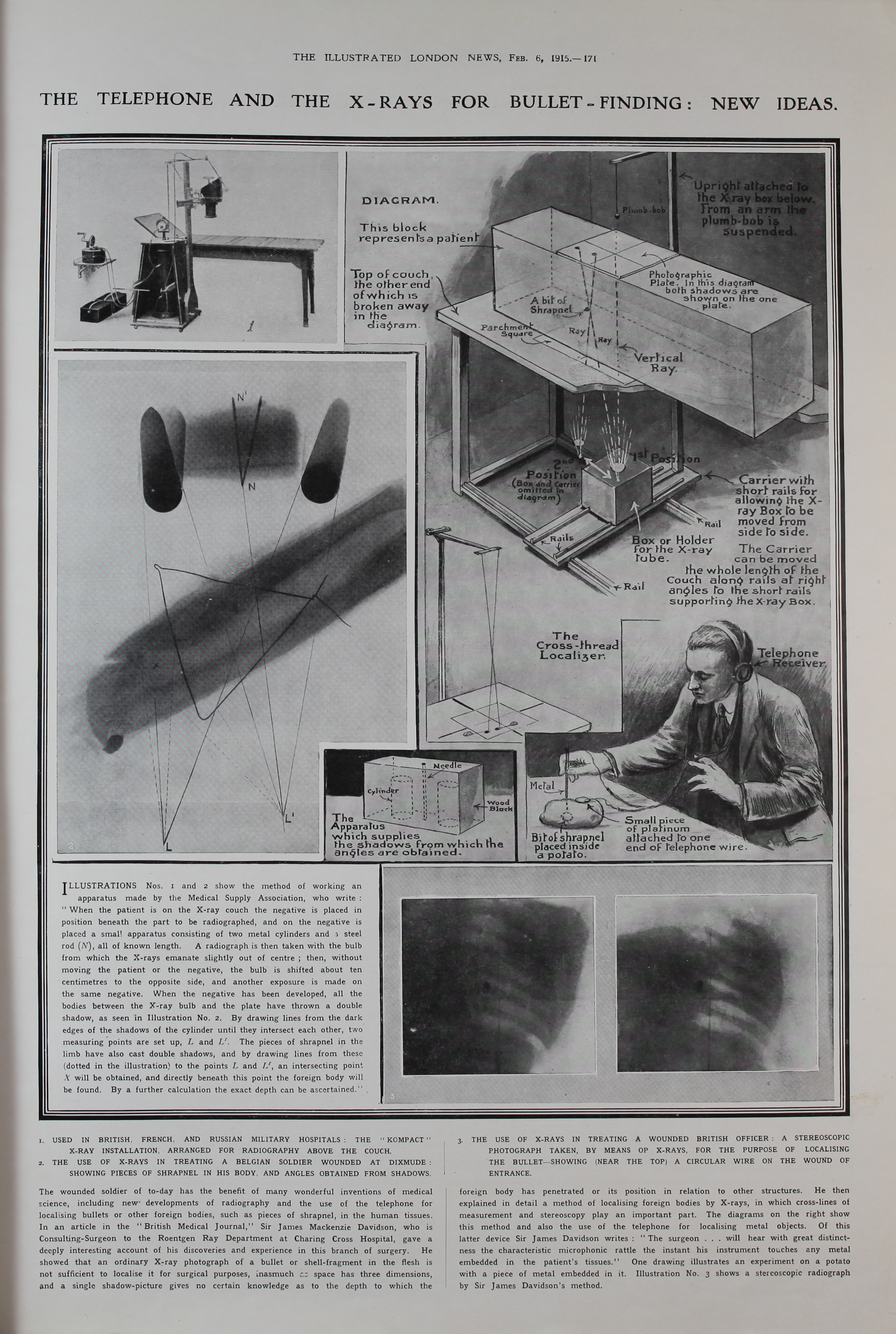
Illustrations Nos.1 and 2 show the method of working an apparatus made by the Medical Supply Association, who write: ‘When the patient is on the X-ray couch the negative is placed in position beneath the part to be radiographed, and on the negative is placed a small apparatus consisting of two metal cylinders and a steel rod (N), all of known length. A radiograph is then taken with the bulb from which the X-rays emanate slightly out of centre; then, without moving the patient or the negative, the bulb is shifted about ten centimetres to the opposite side, and another exposure is made on the same negative. When the negative has been developed, all the bodies between the X-ray bulb and the plate have thrown a double shadow, as seen in Illustration No. 2. By drawing lines from the dark edges of the shadows on the cylinder until they intersect each other, two measuring points are set up, L and L1. The pieces of shrapnel in the limb have also cast double shadows, and by drawing lines from these (dotted in the illustration) to the points L and L1, an intersecting point X will be obtained, and directly beneath this point the foreign body will be found. By a further calculation the exact depth can be ascertained.
- Used in British, French, and Russian military hospitals: the ‘Kompact’ X-ray installation, arranged for radiography above the couch.
- The use of X-Rays in treating a Belgian soldier wounded at dixmude: showing pieces of shrapnel in his body, and angles obtained from shadows.
- The use of X-Rays in treating a wounded British officer: a stereoscopic photograph taken, by means of x-rays, for the purpose of localising the bullet – showing (near the top) a circular wire on the wound of entrance.
The wounded solider of to-day has the benefit of many wonderful inventions of medical science, including new developments of radiography and the use of the telephone for localising bullets of other foreign bodies, such as pieces of shrapnel, in the human tissues. In an article in the ‘British Medical Journal,’ Sir James Mackenzie Davidson, who is consulting-Surgeon to the Roentgen Ray Department at Charing Cross Hospital, gave a deeply interesting account of his discoveries and experience in this branch of surgery. He showed that an ordinary X-ray photograph of a bullet or shell-fragment in the flesh is not sufficient to localise it for surgical purposes, inasmuch as space has three dimensions, and a single shadow-picture gives no certain knowledge as to the depth to which the foreign body has penetrated or its position in relation to other structures. He then explained in detail a method of localising foreign bodies by X-rays, in which cross-lines of measurement and stereoscopy play an important part. The diagrams on the right show this method and also the use of the telephone for localising metal objects. Of this latter device Sir James Davidson writes: ‘The surgeon… will hear with great distinctness the characteristic microphonic rattle the instant his instrument touches any metal embedded in the patient’s tissues.’ One drawing illustrates an experiment on a potato with a piece of metal embedded in it. Illustration no. 3 shows a stereoscopic radiograph by Sir James Davidson’s method.