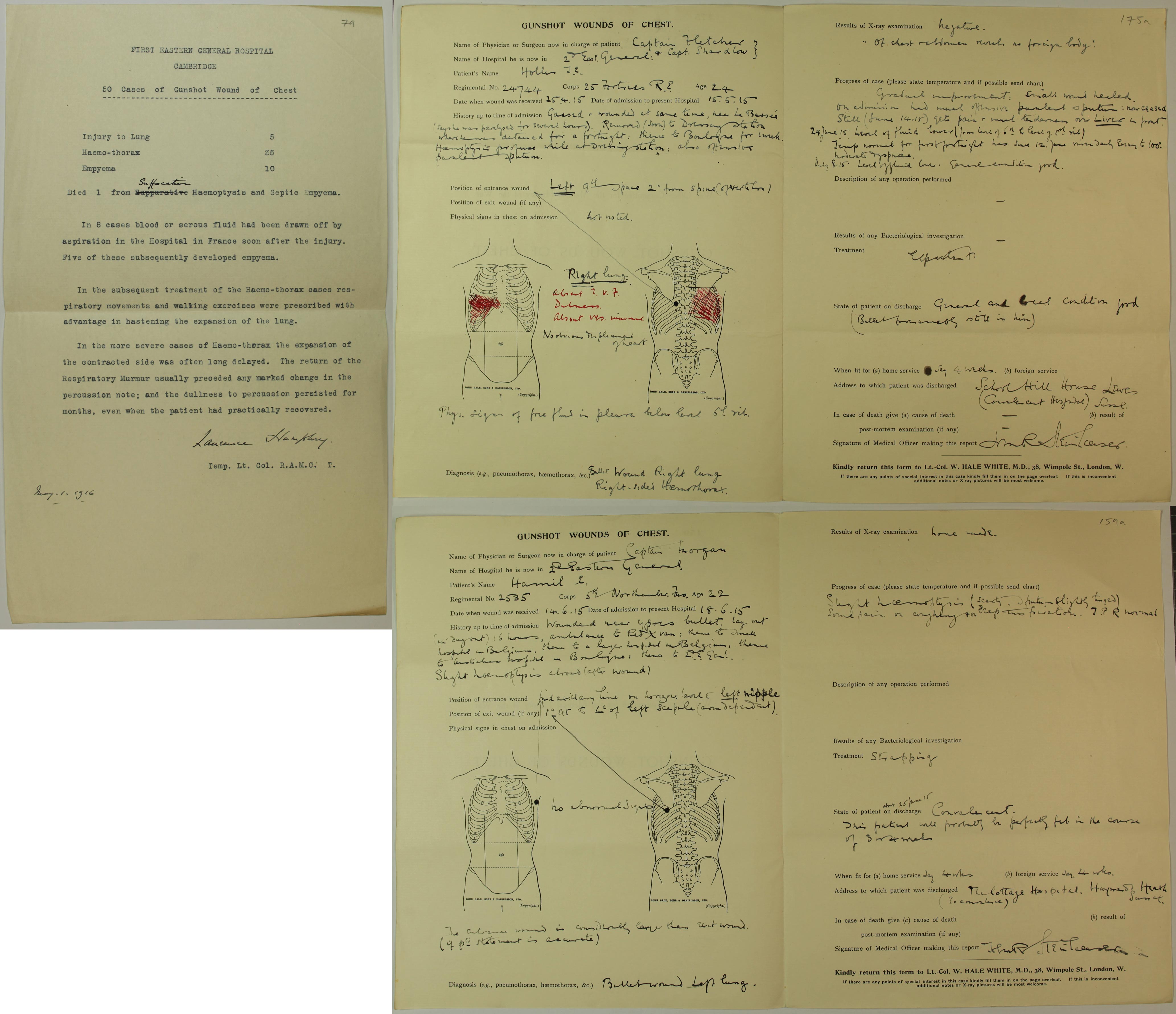
An example of an attempt to compile statistics for gunshot wounds at the First Eastern General Hospital in May 1916, and two cases of gunshot wounds to the chest, (Catalogue ref: MH 106/2115)
Transcript
FIRST EASTERN GENERAL HOSPITAL
CAMBRIDGE
50 Cases of Gunshot Wound of Chest
Injury to Lung 5
Haemo-thorax [blood accumulates in the pleural cavity. This excess fluid can interfere with normal breathing by limiting the expansion of the lungs] 35
Empyema [pockets of pus collected inside a body cavity. They can form if a bacterial infection is left untreated] 10
Died. 1 from suffocation Haemoptysis [coughing blood] and septic Empyema
In 8 cases blood or serous fluid had been drawn off by aspiration in the Hospital in France soon after the injury. Five of these subsequently developed empyema.
In the subsequent treatment of Haemo-thorax [blood collecting between chest wall and your lungs] cases respiratory movements and walking exercises were prescribed with advantage in hastening the expansion of the lung.
In the more severe cases of Haemo-thorax the expansion of the contracted side was often delayed. The return of the Respiratory murmur usually preceded any marked change in the percussion note; and the dullness to percussion persisted for months, even when the patient has practically recovered.
Laurence Humphrey
Temp. Lt. Col. R.A.M. C. T
May 1 1916
GUNSHOT WOUNDS OF CHEST
Name of Physician or Surgeon now in charge of patient Captain Fletcher & Capt. Shardlow
Name of Hospital he is now in 2nd East General
Patient’s Name Hollis J.E.
Regimental No. 24744 Corps 25 Fortress Royal Engineers Age 24
Date when wound was received 25.4.15 Admission to present Hospital: 15.5.15
History up to time of admission
Gassed & wounded at same time, near Le Bassée (he says he was paralysed for several hours) removed (soon) to Dressing Station where he was detained for a fortnight: thence to Boulogne for 1 week. Haemoptysis [coughing up of blood] profuse while at Dressing Station: also offensive purulent sputum [pus & mucus]
Position of entrance of wound Left 9th space 2 inches from spine (of vertebra)
Physical signs in chest on admission not noted
[Two diagram of chest to show wound sites for entrance and exit of bullet(s)]
Right Lung
….
No obvious displacement of heart
Physical signs of free fluid in pleura below level of 6th rib.
Diagnosis (e.g. pneumothorax, haemothorax [blood collecting between chest wall and your lungs]
Bullet wound Right lung. Right sided haemothorax
Results of X-ray examination – negative “of chest & abdomen reveals no foreign body”
Progress of case (please state temperature and if possible send chart
Gradual improvement: small wound healed. On admission had much offensive purulent sputum: now ceased. Still (June 14.15) gets pain & small tenderness over liver in front. 24th June 15, level of fluid lower (from level of 6th rib to level of 8th rib). Temperature normal for the first fortnight, has since 12 June, risen daily & rising to 100 degrees. Moderate dyspnoea [shortness of breath].
July 8th.15 Level of fluid lower. General condition good.
Description of any operation performed –
Results of any Bacteriological examination-
Treatment Expectorant [medicine help expel mucus from the lungs when coughing]
State of patient on discharge- General and real condition good. (Bullet presumably still in him)
When fit for (a) Home service say 4 weeks (b)foreign service
Address to which patient was discharged School Hill House, Lewes Sussex (Convalescent Hospital)
In case of death give (a) cause of death
Post-mortem examination (if any)
Signature of Medical Officer making this report….
GUNSHOT WOUNDS OF CHEST
Name of Physician or Surgeon now in charge of patient Captain Morgan
Name of Hospital he is now in 2nd Eastern General
Patient’s Name Hamil E.
Regimental No. 2535 Corps 5th Northumberland Fusiliers Age 22
Date when wound was received 14.6.15 Admission to present Hospital: 18.6.15
History up to time of admission
Wounded near Ypres, bullet, lay out (in dugout) 16 hours, ambulance to Red Cross van, thence to small hospital in Belgium, thence to a larger hospital in Belgium, thence to Australian hospital in Boulogne, thence to 2nd Eastern General [hospital]. Slight haemoptysis [coughing up of blood] abroad (after wound).
Position of entrance of wound
Mid axillary line on horizon. Level E (entrance] left nipple
Position of exit of wound (if any) 1 inch 4r [4th rib) to left of left scapula… no abnormal signs
Physical signs in chest on admission
[Two diagram of chest to show wound sites for entrance and exit of bullet(s)]
The entrance wound is considerably larger than the exit wound (if patient statement is accurate)
Diagnosis (e.g. pneumothorax, haemothorax [blood collecting between chest wall and your lungs]
Bullet wound Left lung
Results of X-ray examination none made
Progress of case (please state temperature and if possible send chart
Slight haemoptysis (scanty, sputum slightly tinged [with blood]). Some pain on coughing & on deep inspiration. T.P.R. [temperature, pulse, respiration]
Description of any operation performed
Results of any Bacteriological examination
Treatment Strapping
State of patient on discharge- about 25th June ’15, Convalescent. This patient will probably be perfectly fit in the course of 3 or 4 weeks.
When fit for (a) Home service (b) foreign service: say 4 weeks.
Address to which patient was discharged The Cottage Hospital. Haywards Heath Sussex (to convalesce)
In case of death give (a) cause of death
Post-mortem examination (if any)
Signature of Medical Officer making this report….
